

Skeletons and Silicon
Every year, the Hammer Museum offers a unique opportunity for UCLA graduate students to curate an exhibition from the Grunwald Collection of Graphic Arts housed in the Grunwald Study Center. This year, history of science doctoral candidates Marissa Petrou and Twyla Ruby collaborated in organizing a compelling exhibition highlighting the entwined histories of science and art. Skeletons and Silicon: The Art and Science of The Human Bodywill be on view in the Grunwald Study Center through August 2013. Below is their exhibition essay.
A skeletal figure dances gracefully across a celestial field in Nancy Sutor's photomontage Skeleton (1982). Here, the scientific icon has been reimagined and imbued with vitality. In Barbara Morgan's Inner and Outer Man (1972) a luminous x-ray of a skull and spine is superimposed against the dark silhouette of a man's head. In this dual portrait of her recently deceased husband, the two representations comprise a palimpsest, as if past and present are in dialogue with one another. Morgan demonstrates that artists can represent the dynamic qualities of human life through the combination of artistic and medical visual techniques. Sutor and Morgan participate in a broader critique of modern medicine's picture of the human body as static, lifeless and typically masculine. They engage with scientific technologies, materials, and methods to emphasize the vitality and dynamism of the living body. This undertaking in the art community is not new: Sutor's dancing skeleton echoes the gesticulating flayed male in Domenico Barbiere's Skeletons and Ecorches (16th century). This exhibit explores the ways artists have critiqued scientists' representation of the inanimate human form from the Renaissance until the present.
In Riverside Millineries (2001), Deborah Puretz appropriates the silicon wafers of microchips to unite two art-making processes that are often diametrically opposed: traditional printmaking and computer technology. Having etched and inked the wafers for use in print, she layers their impressions with repurposed photos of two turn-of-the century milliners. Puretz refers to the wafers as metaphors of memory which reflect the circuitous paths of the human mind as it processes, stores, and retrieves information. The collage evokes the century-long history of women as computers (in the original sense of one who computes) to attack the stereotype that women are not technically minded. The emergence of modern medicine in the nineteenth century, and its image of the human body, played an important role in defining who may participate in science. Often, the results of medical science were used to argue that women, the poor, and the non-white had a deviant ‘nature’ that rendered them incapable of employing scientific concepts or methods. Puretz, like Morgan and Sutor, refutes these claims by repurposing and mixing techno-medical materials. If we take a long view, we see that this kind of critical exposure of the medical community's blindspots existed even in the 18th century.

Giovanni Piranesi’s The Skeletons (1750-1778) appropriates and re-purposes medical iconography to assert the status of his own artistic knowledge of human nature. The skeleton in the foreground draws heavily from standard anatomical representations, yet here these familiar images are embedded into an allegorical setting. In the sky, the image of the scorpion provides a unifying theme. In medical astrological belief, still prevalent in the 18th century, the scorpion connoted the capacity for reproduction. Reigning over the composition of skeletons, architectural ruins, and lush plants, the scorpion suggests that the print is about organic and social regeneration, an aspect of the body that anatomists were unable to represent.
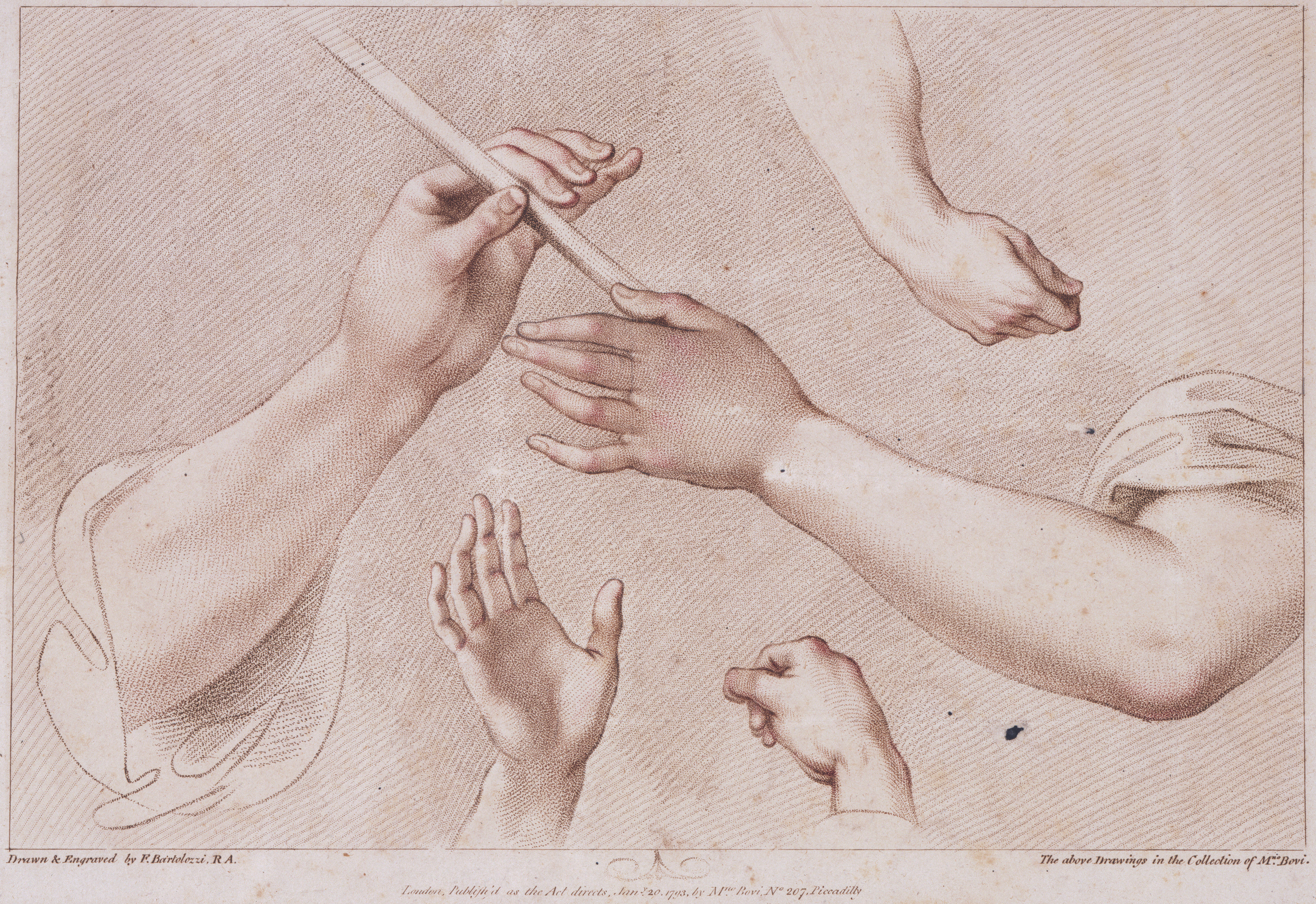
Like his contemporary Piranesi, Francisco Bartolozzi departed from the traditional line engravings used to illustrate anatomy texts to find a more accurate means of depicting the vitality of the human body. Bartolozzi developed the stipple engraving technique to convey movement through complex shadows and tone. This engraving serves as a technical manual that advertises the refinement of an earlier technique. In the image of multiple disembodied arms, where the engraver’s arm renders similar specimens as two-dimensional images, Bartolozzi demonstrates that the skills of the artist are a type of knowledge based in the muscular movements of the hand. The stipple engraving makes a claim for the importance of invention and embodied knowledge in investigations of nature and the human body.
Fritz Faiss' 1933 portrait of 16th century German physician Paracelsus evokes the emblematic figure of a modern-day scientist, pictured in his laboratory among beakers, distillators, and other instruments of experimentation. This same equipment was used by Paracelsus in his alchemical investigations into the ongoing processes of transmutation, generation and decomposition. In his own day, Paracelsus was expelled from the elite medical community for rejecting text-based knowledge in favor of concrete experimentation.
Faiss, himself both a German physician and an artist, compares his 20th century work in laboratory medicine to the ancient, holistic philosophy of alchemy. For Faiss, as for all the artists in this exhibit, experimentation was integral to representing both the body and the forces that animate it. Each artist questions the conservatism of the medical community and the limited knowledge represented in static images of the human form. Yet these artists also appreciate the experimental possibilities made available through medical skills, materials and techniques. The solution they propose is the collaboration of art and science.
Skeletons and Silicon: The Art and Science of the Human Body
Curated by Marissa Petrou and Twyla Ruby
On view starting June 28, 2013
Grunwald Study Center, Hammer Museum
Hours: M-F 9am-5pm
Call 310-443-7078 for an appointment to view the exhibition.
About the Curators
Marissa Petrou is a doctoral candidate in the history of science at UCLA. Her dissertation explores how scientists in Imperial Germany utilized museums to develop the new science of ethnology via text, image and object. Twyla Ruby is a graduate student in the history of science at UCLA interested in the visual culture of science in the early modern period. Marissa and Twyla have previously curated history of science exhibits at the UCLA research libraries.
Captions
Top to bottom:
Domenico del Barbiere, Skeletons and Ecorches, c. 1540 - 1545. Engraving, 9 3/8 x 13 1/4 in. UCLA Grunwald Center for the Graphic Arts, Hammer Museum.
Deborah Puretz, Riverside Millineries, 2001. Etched inked wafers (slices of silicon, later cut apart as computer chips) collaged with found documents and gelatin silver prints, 15 5/8 x 15 5/8 in. UCLA Grunwald Center for the Graphic Arts, Hammer Museum. Courtesy of the artist.
Francesco Bartolozzi, Untitled, 1793. Stipple engraving, 8 11/16 x 12 11/16 in. UCLA Grunwald Center for the Graphic Arts, Hammer Museum.

